About the dataset
This dataset comprises of contrast-enhanced computed tomography (CECT) images of the abdominal region, featuring 160 patients diagnosed with various ailments, such as hepatocellular carcinoma (HCC), intrahepatic cholangiocarcinoma (ICC), and liver hemangioma. These images were acquired using diverse CT scanners, following the administration of an iodine contrast agent at a weight-appropriate dosage (1-1.5 ml/kg) prior to the examination.
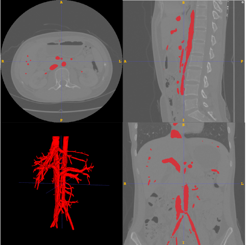
During the meticulous process of annotating the liver and intrahepatic space-occupying lesions, we carried out precise delineation of the hepatic vasculature, encompassing the hepatic artery, portal veins, and hepatic veins, with special emphasis on the portal veins. This comprehensive annotation enables a deeper understanding of the anatomical relationship between these lesions and the intricate network of blood vessels within the liver. The rigorous manual labeling of these structures facilitates accurate segmentation of liver segments by aligning with the vascular architecture.
The intricate pathways of intrahepatic blood vessels serve as the foundational basis for devising liver resection surgical plans. The precise annotation of liver blood vessels in this dataset holds significant utility for the development and validation of models aimed at predicting resection volume and helping with surgical planning. Such models can greatly benefit clinical practice by offering invaluable insights into surgical decision-making and outcomes.
Access Guideline
